
The Body of Bubbles: Necropsy Report
WHITE SANDS RESEARCH CENTER
Pathology Report
Necropsy
ID No. CA1376
Sex: Male
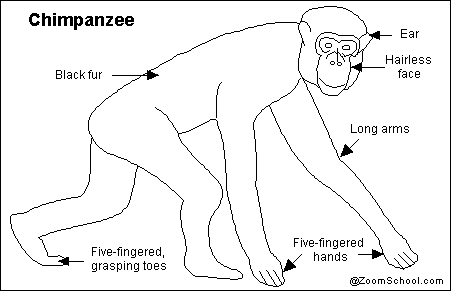
Species: Chimpanzee
Date of Death: 29 June 2015
Pathologists: Robert Mason, DVM and Gary Silbert, DVM, Ph.D.
GROSS EXAMINATION:
Dr. Frost called Dr. Mason approximately 10:30 a.m. 29 June 2015 with message that a chimpanzee was found dead and that it “appeared like bloat, the abdomen was reddish purple.”
No additional clinical data was provided with the animal.
The animal was taken from the 40 degree F morgue where it was placed on the necropsy table at approximately 1:30 p.m. June 29, 2015.
The tatoo on the animal’s inner right thigh was 1376.
The animal was a young adult male in good body flesh and hair coat. The animal was in rigor mortis. The mucous membranes of the mouth were a dark purple color (cyanotic). No oral lesions were present. The lower abdomen had liver mortis suggesting ventral recumbency post mortem. The skeleton appeared normal except for an occipital bone asymmetry. No signs of trauma were found externally.
A standard “Y” incision was made and the subcutaneous were examined and no lesions were found. The abdominal and thoracic viscera were examined and the stomach contained watery contents with no solid ingesta. The gastricmucosa and valves appeared normal. The entire digestive tract was examined and no lesions were found. The urinary bladder contained approximately 150ml of clear urine. The kidneys and urinary tract appeared normal. The adrenals, pancreas, spleen, and other abdominal tissues appeared normal.
The heart had multiple petechial hemorrhages on the epicardium especially the right ventricle. No other abnormalities were seen in the heart or associated great vessels.
The lungs did not collapse completely and diffuse red hepatization was in all lobes. The bronchial tree had thick cloudy (purulent exudate) tenacious fluid filling the lumens. Similar exudate was adhered to the lower tracheal wall. The structures of the oral cavity and pharynx appeared normal.
The cranial cavity was opened and the meninges over the entire brain was cloudy with a purulent exudate. The meninges of the spinal cord had a similar appearance.
Bacterial cultures were taken of the meninges and bronchi. Samples representative of all organ systems were collected in buffered formalin.
Gross Diagnoses:
Pneumonia, acute, suppurative, diffuse, all lobes of the lung, etiology_Streptococcus pneumoniae_.
Meningitis, suppurative, diffuse, meninges of brain and spinal cord, etiology_Streptococcus pneumoniae.
Comments:
The presence of suppurative meningitis with pneumonia is most commonly caused by pyogenic bacteria but can also be caused by mycoplasma and other less common organisms. Samples of brain was frozen at -70 C for additional etiologic study if the bacterial cultures and histological examination of collected tissues do not result in isolation and identification of a probable pathogen. Tests were also ordered of any isolated pathogens.
Note:
_Streptococcus pneumoniae_ subsequently isolated from the bronchus and brain and had identical antibiotic sensitivity profiles suggesting high probability that the animal had a primary pneumonia that extended to meninges.
Male Genital System: The male genitalia system is unremarkable.
Robert Mason, DVM, Gary Silbert, DVM, Ph.D.
Hi Carlton-
I think this is a great idea for a performance, but I’m a bit confused. Is this the actual text or did you manipulate it in some way? Perhaps you’re suggesting it could be read and performed? Are you posting it so you can perform next week? I need a bit more context here I think! Thank you! -Amanda
Yes, I have manipulated and appropriated as necessary for creative effect. I plan to perform the piece in class.
Should I know Bubbles?